Please note: Information in this article is correct at date of publication, 16 January 2013.
Identifying the composition of both objects needing conservation and the materials used in their conservation is an important facet of the treatment process. Proper identification can elucidate the original state of an object, the nature of degradation present, and the possible effects of different conservation treatments on the object. Students at West Dean are therefore fortunate to have the opportunity to learn how to make use of the some of the different analytical equipment available and useful to conservators in our modest but growing analytical laboratory.
What are West Dean's analytical capabilities?
One of the most frequently used instruments is our Fourier-Transform infrared spectrometer (FT-IR), which uses infrared light in its analysis. It gives particularly good results with organic materials, and works by measuring the amount of infrared transmitted or absorbed by different bonds in a material as a means of identification. Our current apparatus is an older model but following a successful grant application, a new instrument will be arriving later this month with advanced capability and an easier method of taking samples.
West Dean also employs a fluorescence microscope, looking at surfaces with different parts of the visible and UV spectrum in order to identify materials according to the color of their fluorescence; a visible light spectrophotometer, useful for measuring surface color readings, and which can aid in color change measurements; in addition to a UV-Vis spectrometer that takes spectra of solutions, and is also helpful in evaluating color change through the observation of the UV range.
Our other oft-used piece of tech is our handheld X-ray fluorescence spectrometer, which will be discussed in greater detail below as recent developments have brought it into prominence.
X-ray Fluorescence Spectrometry (XRF)
How does XRF work? It's all down to the radiation. X-rays, along with gamma and ultraviolet radiation, form part of the electromagnetic spectrum known as ionizing radiation. When a material is hit with ionizing radiation, electrons are ejected from the constituent atoms, and the strength of this radiation can be sufficient to dislodge electrons from the inner shells of an atom. In this case, the empty spaces left by the missing electrons render the atoms unstable, and so electrons from higher orbitals fall into those places, losing energy as they do and emitting it in the form of radiation, hence the "fluorescence" part of the term XRF. This fluorescent energy is what the instrument detects, and is characteristic to each element: the difference in energy between each orbital is discrete or quantized and known to science. XRF works particularly well with inorganics and is extremely useful in making qualitative descriptions of metallic materials.
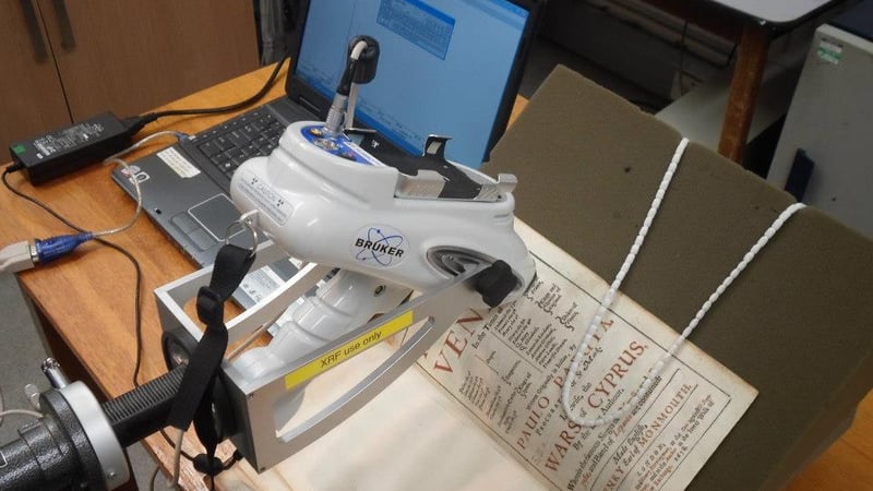
As one can imagine, X-rays and other ionizing radiation can be dangerous. Changing the elemental make-up of something will of course have consequences, and as is evident today, radiation poses a significant problem and often leads to cancer and other types of radiation-related illness. With this in mind and as part of new regulations, staff and students underwent radiation protection supervisor training to ensure proper precautions are taken when using radiation as an analytical tool.
Radiation exposure and analysis
During the training we discussed several different important factors to consider in employing radiation for analysis, including the legal and scientific aspects of its use. For comparison purposes, our radiation safety advisor showed us radiation amounts for some of the common circumstances in which the average person might encounter radiation.
[The SI unit for radiation is the Sievert (Sv), a large quantity of radiation exposure, and so for most dosages the amount of radiation is given in milliSieverts (mSv), one-thousandth of a Sievert, or in microSieverts (μSv), one-millionth of a Sievert.]
For example, a dental X-ray will subject a person to between 5 and 10 μSv; hence the reason that a patient is given a lead apron to wear and the technician leaves the room. A CT scan gives off about 0.01 Sv. Symptoms of radiation sickness begin to appear anywhere from 0.75 Sv to 1.0 Sv. By comparison, a handheld XRF instrument will produce about 1 μSv per hour, a tiny amount compared to other sources of radiation. On top of that, most exposure times necessary for XRF identification are counted in seconds, further minimizing the amount of radiation. However, it's important to remember that radiation poisoning is cumulative. Just because the dose is small doesn't mean that unnecessary exposure won't be detrimental. Proper precautions are absolutely essential to maintaining low levels of exposure to those concerned, which reflects an industry policy known as ALARP: As Low As Reasonably Practicable.
ALARP takes as its core principle in practice that unnecessary exposure should be absolutely minimal. There will be obviously be some exposure to the analyst, but this is an accepted risk that the analyst chooses to take in order to conduct the testing (hence the word "practicable"). But exposure to others should be eliminated insofar as it is possible, and with our radiation safety advisor, we learned how to do so.
Minimizing unnecessary exposure and creating safe analytical environments
One of the most important takeaways from this training was a more precise understanding of the penetrative abilities of X-rays, which is essential for creating a safe analytical environment. An average brick (in terms of density) actually absorbs 99.9999% of X-radiation, the same as 1 mm of lead. Another brick or 1 mm of lead will absorb another 99.9999% of the remaining radiation, leaving just one hundred-millionth of the original. It's a good thing we wear lead aprons at the dentist!
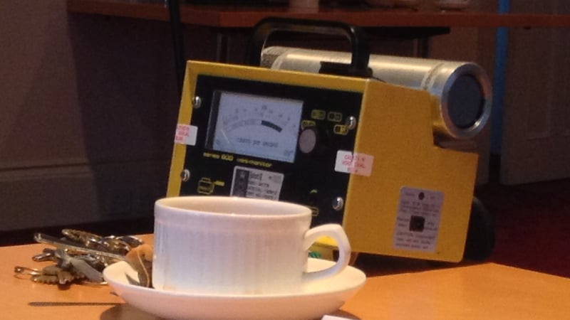
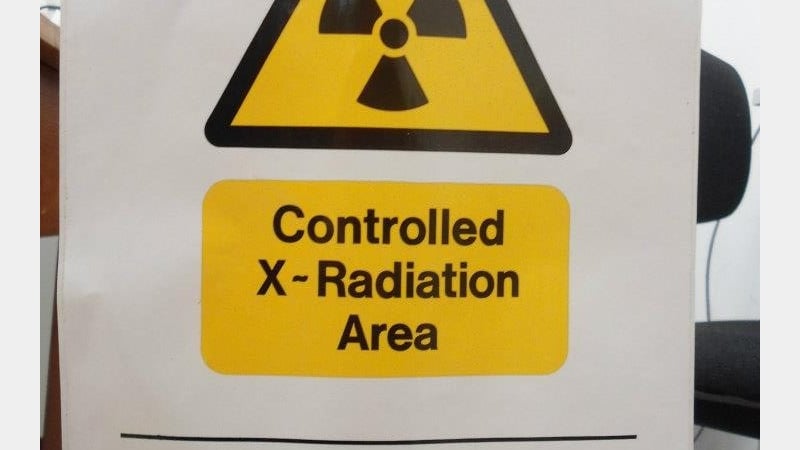
Establishing a proper area for conducting analysis is also important. It's important to think about not just those present in a workshop or studio but also about those who might be on the other side of a wall or out a window. A thick wall will do the trick (think of the two bricks), and to be safe, one that doesn't have any other people on the opposite side. An adequate perimeter should also be established with proper signage informing others that radiation is being used. It was established, using radiation detection equipment, that the advised distance to be kept on either side of a handheld XRF is between four and five meters. This is determined by using the inverse square law, which states that for every unit increase in distance from a source of radiation, the intensity of the radiation decreases accordingly. For example, if the distance from a radiation source is doubled, the intensity is halved.
XRF is an extremely useful analytical tool. Used properly, it can be invaluable in choosing appropriate treatment methods for conservation objects; but like many other tools, used incorrectly, it can be dangerous. Having learned how to put the necessary precautions into practice, students at West Dean are capable of safely using XRF as a contextual and analytical component of practical work.
